Responsible: Dr. Sofía González and Dr. Jaume Capellades
The Department of Radiology has two CT equipment (a 16-crown siemens somaton sensation team and a GE Healthcare Discovery CT750 HD team) and two Magnetic Resonances (a Philips 3T Achieva 3.0TX Series team and a GE team of 1, 5 T sygna excite).
There are dedicated and specific protocols within the management of patients with epilepsy (Fig. 1).
- Diagnostic MRI for the detection of the possible pathology causing epilepsy.
- Pre-implantation studies of electrodes for the invasive diagnostic processes.
- DTI study prior to implantation of electrodes or surgery.
- Functional MRI: non-invasive study in which the patient performs different tasks or "paradigms" within the resonator. The procedure allows to evaluate functional brain areas that must be preserved during surgery; therefore it is of great help in planning the surgery
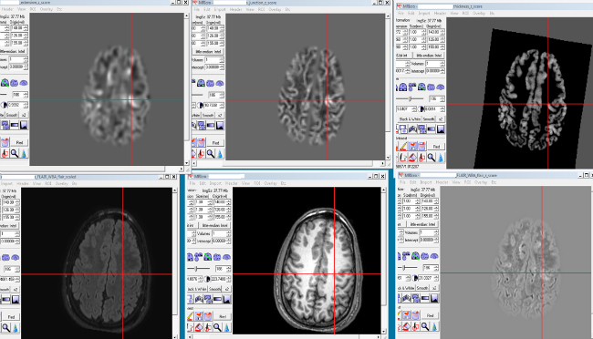
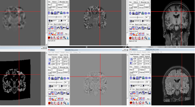
In addition, the MAP software (Morphometric Analysis Program, Huppertz, 2008) has a postprocessing program, adapting the morphometry methodology based on Voxel (VBM) to detect subtle anomalies and help In the process of diagnosis of the epileptogenic focus Fig. 2 and 3).
 Fig.2
Fig.2

Fig.3
Interventional Neuroradiology
Responsible: Dr. Elio Vivas
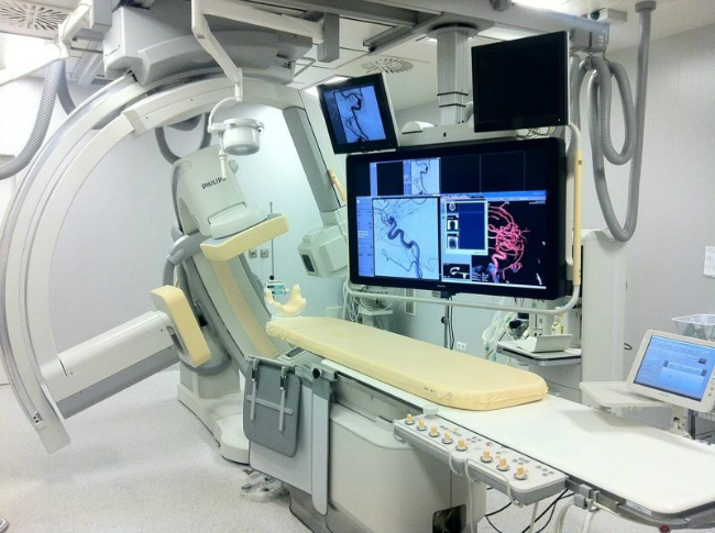
To perform the Wada test, it is necessary to perform carotid digital angiography. These evaluations are also performed for some implantation procedures for intracerebral electrodes. To do this the hospital has a state-of-the-art angiographer Allura Clarity Phillips.

Angiographer Allura Clarity Phillips


